CHICKEN LEG DISSECTION LAB
Introduction:
To identify and describe the structure and function of tendons, ligaments and cartel ge; identify muscles and their function; observe bone and its components; gain experience dissecting fresh biological material.
Procedure:
- CHECK
- Examine the outside skin tissue. Slimy, stretchy, rigid, clumpy and fatty. I noticed the fascicles and the fascia inside the skin tissue.
- When cutting away the skin, I noticed the strong connective tissue, fascia and motor neurons pulling the skin to the muscle.
- Connective tissue appearance.
- Describe the appearance of the connective tissue. The appearance of the connective tissue seems to be translucent, slimy and airy, almost spiderweb like.
- What type of connective tissue is this? This connective tissue is the fascia.
- Observe the yellowish clumps of fat tissue found outside the skin.
a. Describe the fat. The fat is yellowish white, rigid with clumps of structure throughout. It is slimy, stretchy, loose with thick filaments.
b. What are at least two of its functions? Fat cells serve as a layer of insulation for connective tissue as well as a protectant around internal organs and they store energy.
c. Give the biological term for the type of cells that store fat. Adipocytes, also known as lipocytes and fat cells, are the cells that primarily compose adipose tissue, specialized in storing energy as fat.
- Observe bundles of pale pink tissue surrounding the bones. Some muscle tissue and muscle cells are covered with fascia and some without. Without the fascia, the muscle is actually more solid, not as 'shiny'. Sponges, firmness, fibers and ligaments.
- Observe, with you naked eye bundles of muscle tissues surrounding the bones. Separate the bundles of muscles by separating them out with your fingers. Begin by inserting your thumb into the muscle of the lower leg. You will need to push forcefully through the connective tissue covering the muscle, but it will give way at the natural separations between the muscle bundles. Continue separating the muscle into bundles by forcing your thumb and fingers through the muscle until you are able to distinguish several muscle bundles.
- Describe the arrangement of the muscle bundles. Some muscle bundles have more tendons that are denser and some less dense. They seem to be layered on top of one another with connective tissues throughout.
- Do you see just one muscle, or are there many muscles present? There are many muscles present.
- How can you tell? There are a few muscles present because they are separated by connective tissue such as myofibrils, or sacramere and neuromuscular junctions
- Describe any patterns or arrangements you observe such as bundles surrounded by connective
tissue. Each muscle is layer on top of each other with connective tissues surrounding each muscle, tendons in between and nerve cells underneath, connecting the tissue to the bone.
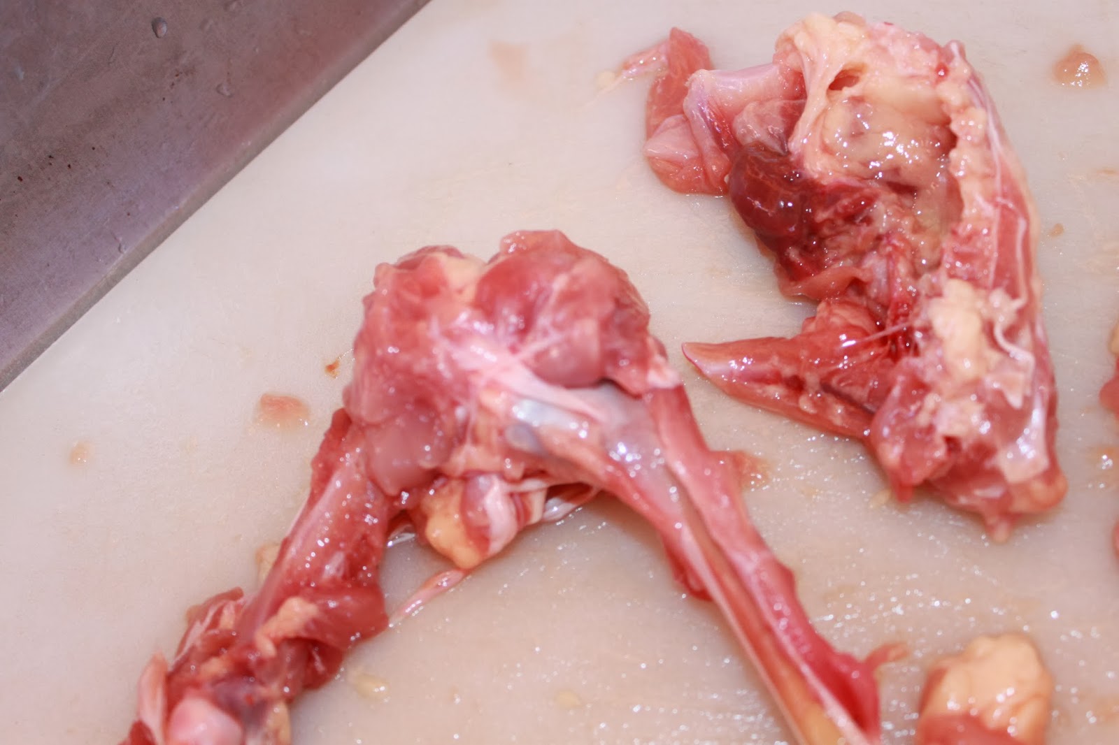
- The strong shiny, white cords, called tendons, hold the muscle to the bones. Some of these tendons will pull away from the bone as you separate the muscle bundles. Find the tendons of the chicken leg. Using the dissection scissors, cut across line A (Figure 1). Observe the numerous tendons and pull the freed muscles down and away from the bone, as if you were peeling a banana. Be careful you don’t any ligaments that attach bone to bone. Look closely at the ligaments. Examine the two bones in the lower leg. The large bone (Bone A) is the tibia. The small, toothpick-like bone (Bone B) is the fibula.
- What sort of connective tissue are tendons composed of? Tendons are composed of dense fibrous connective tissue.
- Skeletal muscle function.
- What are skeletal muscles? Skeletal muscles are muscles composed of cylindrical multinucleate cells with obvious striations. Skeletal muscles attach to the body's skeleton.
- What are their function? Skeletal muscles interact with the skeleton and correlate with the nerves to cause the bones to contract and relax.
- 11. Remove a single muscle by cutting the tendons and peeling the muscle away from the bone. Nerves are generally thin, threadlike white strands found between the muscle and the nearest bone. Look for the nerve specimen. The texture is much different from a tendon or bone. It is rather slippery.
- a. Did you find it? Yes, I did.
- 12. Remove the muscle that covers Joint B by cutting parallel to the femur, upward toward the backbone. Remove pink muscle tissue until you see a shiny white sheet of ligament that covers the joint. Present is an exterior ligament that holds the femur in the hip socket.
- a. What type of connective tissue composes the ligaments? The ligaments that holds this joint together is dense fibrous connective tissue.
- 13. Remove all remaining muscle to expose the bones of the chicken leg.
- 14. Cut onto the hinge joint by cutting into the top of the covering of the joint from the femur side. It will become apparent that you must remove the kneecap area to expose the menisci and ligaments within. Pull up on the kneecap area and cut through it with scissors. You will have cut through the bursa, a sac that acts as a shock absorber for the knee joint. These are found in every joint.
- 15. Pull the covering back and look inside the joint. You will see more white bands of ligaments holding the bones together. Observe the shiny, white layer covering the ends of the bones is cartilage. It helps the bones slide smoothly when the leg bends.
- 16. Bend the specimen at Joint B (Figure 1) and rotate the femur in all directions. Refer to pages 113-115 in your textbook and answer these questions.
- a. What type of joint is this? Joint B is a synovial ball.
- b. What type of movement is being demonstrated? The movement being demonstrated is known as circumduction.
- 17. Cut the ligaments at the joint between the upper and lower leg. Examine how the bones fit into each other.
- a. Describe the texture of the ends of the bones at the joint. The texture of the ends of the bones at the joint are smooth and connective.
- b. What is the specific name of this type of connective tissue? The specific name of the type of tissue found at the joints is cartilage.
- c. What occurs when the cartilage at joints wears away? When the cartilage at the joints wears away, this is called Osteoarthritis.
- d. How is a joint of this type built to delay this event? Because tendons, muscle and tissue surround the joint , the joint is built to delay this event. As long as you get enough calcium and vitamin D and continue to exercise, you could prevent this problem.
- 18. If you could see inside the bone.
- a. What soft material would you find? If you could see inside the bone you would find bone marrow.
- b. Name three specific types of cells present here. The three bone cells that are present are osteoblasts, osteocytes and osteoclasts.
- c. Name the three functions of bone. Three functions of the bone are to protect vital organs, structure and movement.
- 19. CHECK
- 20. FINISHED.
- CONCLUSION.
- Although it was quite nauseating to do this lab (possibly because I am a vegetarian), it was definitely interesting to discover how similar a raw chicken leg is to our own. All of the tendons, connective tissues, muscles, bones that nerves that are present work together to create contractions and relaxations in our body's movement (and the chicken's) everyday.